Menu
In Vivo Small Animal Imager (IVIS Spectrum, Xenogen)
IVIS Spectrum, the small animal imager from Xenogen, Perkin Elmer, USA is an advanced animal imager that can support both fluorescence and bioluminescence imaging with high sensitivity. This equipment supports both Trans and epi illumination enabling 3D diffuse fluorescence tomography for depth perception. Also equipped with small animal anesthesia unit.
Location: Main Campus
Contact:
Saravana Kumar M
msaravana@rgcb.res.in
Flow Cytometer Sorters
High Speed Flowcytometer Sorter System: (FACSAria III)
FACSAria III is a Bench top fixed aligned high speed 4 way sorter system from Becton Dickinson, USA and is equipped with the laser lines, 488 nm, 355 UV, 561, 405 Violet lasers and 633 nm. The machine allows plate sorting. Also equipped with aerosol management system.
Location: RGCB Jagathi Campus
Contact:Indu Ramachandran,
indur@rgcb.res.in
91-471-2529-507, 9447146937
High Speed Flowcytometer Sorter System: (FACSAria III)
FACS Aria III, Bench top fixed aligned flow cytometer is a high speed 4 way sorter system from Becton Dickinson, USA and is equipped with the following laser lines 488 nm Laser, 445 nm, 405 nm Violet lasers, 561nm laser and 633 nm laser.
Location: RGCB Akkulam BIC
Contact:Surabhi S V,
surabhi@rgcb.res.in
91-471-2764-081, 9633999424
Beckman Coulter Astrios EQ High Speed Cell Sorter
Six-way jet-in-air sorter with 7 lasers (355nm, 405nm, 488nm,532nm, 561nm, 592nm,640nm) and 22 fluorescent color capability. It has dual forward scatter PMTs that allow researchers to analyze biological samples ranging in size from 0.2um to 30um. The instrument is capable of simultaneous 1-6 way sorting and is equipped with Cyclone sorting for single cell sorting. Available nozzle sizes are 70um and 100um. It is contained within a Baker SterilGARD Class II biological safety cabinet for optimised sterile sorting.
Location: RGCB Akkulam BIC
Contact:Surabhi S V,
surabhi@rgcb.res.in
91-471-2764-081, 9633999424
Confocal Microscopy
Confocal Laser scanning Microscope with high sensitivity spectral detector (Olympus FV3000)
Olympus FV3000 is equipped with a high sensitivity spectral detector (HSD) with GsAsP PMTs which enables it to view samples having weak emission. The desired emission range can be selected using the spectral detectors. The Diode Laser lines available are 405nm, 488nm, 514nm, 561nm and 640nm with 4x, 10x, 20x, 40x and 60x objectives.
Location: RGCB Jagathi Campus
Contact:Ciji Varghese , L.Parvathy, Athira S S
cijiv@rgcb.res.in,parvathyl@rgcb.in,athirass@rgcb.res.in
91-471-2529-439
Confocal Laser Scanning Microscope with GaAsP Detector for Multifluorescence and Live Cell Imaging (Leica SP8 WLL Confocal Microscope)
Leica SP8 Spectral Confocal with WLL is an advanced confocal microscope from Leica Microsystems, Germany. This equipment is configured with white light laser (WLL) that can support any laser lines between 470-670nm and AOBS for filter less emission tuning, in addition to the highly sensitive GaAsP detector.
Location: RGCB Jagathi Campus
Contact:Ciji Varghese, Ayswarya R S
cijiv@rgcb.res.in, ayswaryars@rgcb.res.in
91-471-2529-439
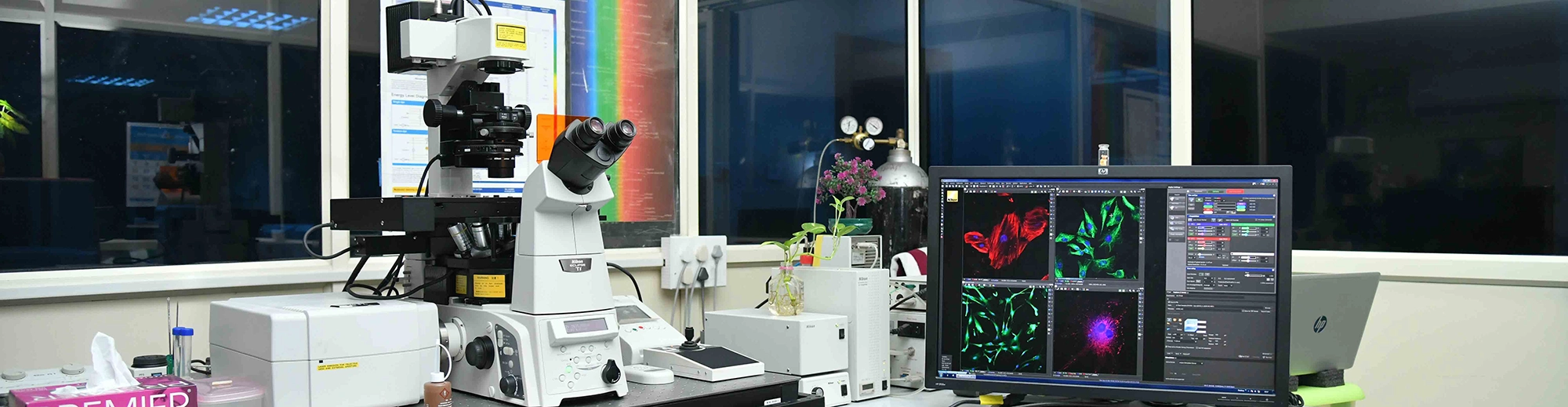
Multiphoton Confocal Imaging system: Nikon A1R MP
The multiphoton confocal imaging system A1R MP is the dual line IR laser integrated multiphoton confocal with all required accessories and imaging softwares from Nikon, Japan. The system is capable of simultaneous or sequential image collection using galvanometer and resonant scanners and four photomultiplier fluorescence detectors and a DIC transmission detector. The multiphoton component is equipped with the following laser excitation source detectors in addition to the conventional visible detectors.
- Coherent Chameleon II Dual Line Tunable laser for MP imaging with a tuning range of 660nm to 1320nm as output A and 1040nm as output B.
- Galvanometer and Resonant scanners.
- Nikon Apo 25xW, CFI Apo 40xW NIR and CFI 60XW NIR objective for MP imaging.
- Four PMT detectors and four GaAsP NDD (reflected light) detectors.
- NIS Elements software controlling all microscope components.
Location: RGCB Akkulam BIC
Contact:Arun Jyothi
9656040132
High Content Imaging System: Imagexpress Confocal HT.AI, Molecular device
The ImageXpress® Confocal HT.ai High-Content Imaging System is an advanced imaging platform with seven-channel laser light source and eight imaging channels to enable highly multiplexed assays while maintaining high throughput by using shortened exposure times. Designed for experiments involving live cells as the spinning disk reduces phototoxicity and enables fast acquisition. The system supports slides and one to 1536-well microplates, round or flat bottom, low to high profile.
- Inverted microscope (ImageXpress Confocal HT.ai)
- Precision motorized X-Y (sample) stage
- Dual disk unit allowing selection between 3 modes
- 60 micron pinhole disk for basic confocal requirements
- 50 micron slit for high throughput confocal requirements
- Widefield (not-confocal) imaging mode
- 4 megaPixel sCMOS
- 8 position emission wheel and 5 position dichroic filter wheel
- Temp and air control system
Location: RGCB Akkulam BIC
Contact:Laiza Paul,
laizapaul@rgcb.res.in
91-471-2764-046
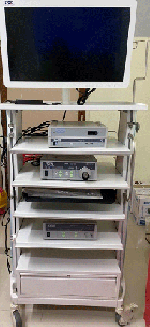
Rodent Endoscopy System
Mouse colonoscope system: Coloview high-resolution mouse endoscopy system (KARL STORZ SE & Co. KG, Germany). This system can be used for colonoscopic examination and biopsy in mice and rats.
Location: RGCB Jagathi Campus
Contact:Dr. Archana S
archanas@rgcb.res.in
91-471-2529-573